
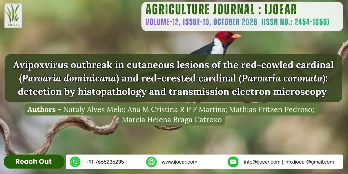
Avipoxvirus outbreak in cutaneous lesions of the red-cowled cardinal (Paroaria dominicana) and red-crested cardinal (Paroaria coronata): detection by histopathology and transmission electron microscopy
Abstract
The red-cowled cardinal (Paroaria dominicana) and red-crested cardinal (Paroaria coronata) are birds of the family Emberizidae naturally found in northeastern and southern Brazil, respectively. These species are targeted by illegal trade because of their colorful plumage and are considered potential viral reservoirs owing to their migratory characteristics. Avian pox is a highly infectious disease caused by poxviruses of the family Poxviridae and genus Avipoxvirus that affects domestic and wild birds. Between January 2006 and September 2007, 72 birds, including 14 red-crested cardinals and 58 red-cowled cardinals, were referred after sudden death by CRAS-PET to the Electron Microscopy Laboratory of the Biological Institute of São Paulo for investigation of viral agents. At necropsy, small cutaneous nodular lesions were observed on the feet of one red-crested cardinal and ten red-cowled cardinals. Some birds had lesions characteristic of avian pox on the feet and/or beaks, consisting of whitish or yellowish nodules and desquamating crusts. Lesion fragments were processed for histopathology and transmission electron microscopy. Light microscopy revealed hyperkeratosis, epithelial hyperplasia, hydropic degeneration, and eosinophilic inclusions corresponding to Bollinger bodies in skin and/or mucosal cells. Negative staining identified numerous typical poxvirus particles in ten crust-suspension samples from red-cowled cardinals and one from a red-crested cardinal. Some particles were enveloped and brick-shaped, with irregularly arranged tubules on the outer membrane, and measured 280 × 260 nm. Ultrathin sections revealed type A intracytoplasmic inclusion bodies, or Bollinger bodies, containing mature or complete oval viral particles measuring 330 × 260 nm, with a biconcave nucleus or dumbbell-shaped internal core. Type B inclusion bodies, immature particles budding from dense amorphous filamentous material, developing particles with spikes on their outer surface, fibrillar inclusions, and deformed nuclei with densely condensed chromatin were also identified. Immunocytochemistry with colloidal gold labeled viral particles and antigenic sites in intracytoplasmic vacuoles and cellular debris. This is the first record of avian pox in the red-crested cardinal (Paroaria coronata) and the second in the red-cowled cardinal (Paroaria dominicana).
Keywords
Download Options
Introduction
The red-cowled cardinal (Paroaria dominicana) (LINNAEUS, 1758) and the red-crested cardinal (Paroaria coronata) (MULLER, 1776) belong to the order Passeriformes and genus Paroaria. The red-cowled cardinal, also known as cabeça-vermelha or cardeal-do-nordeste, is characteristic of the Caatinga and occurs mainly in northeastern Brazil, although it is also found in southern Bahia and Minas Gerais. The red-crested cardinal occurs in Brazil, Uruguay, Paraguay, Bolivia, and Argentina (SICK, 2001).
Both species are frequently affected by the illegal wildlife trade. The red-cowled cardinal has been identified as one of the most commonly traded species in open-air markets in the Recife Metropolitan Region and as the bird most frequently seized in the Campina Grande region, Paraíba (MENEZES et al., 2003; PEREIRA & BRITO, 2005). Of 1,808 birds seized by the São Paulo Department of Parks and Green Areas, 148 were red-cowled cardinals (P. dominicana) (CAVALHEIRO et al., 2003). Red-crested and red-cowled cardinals are also among the species most commonly seized in the South and Central-West regions, while the red-cowled cardinal is among those most commonly seized in southeastern and northeastern Brazil (IBAMA, 2007).
Wild birds are considered potential viral reservoirs because their migratory behavior allows long-distance movements and dissemination of viruses of epidemiological importance. In addition, stressors such as capture, transport, confinement, and dietary or climatic changes may trigger viral processes, leading to sudden outbreaks and varied clinical manifestations (CUBAS & GODOY, 2004).
Diseases affecting avian skin have gained increasing importance because they can cause reduced egg production and growth, blindness, and, in some cases, death. Although natural avian poxvirus infection is better known in domestic poultry, it has also been reported in free-living birds (FENNER et al., 1992).
Avian pox is caused by viruses of the family Poxviridae and genus Avipoxvirus. Poxviruses have double-stranded DNA genomes 130–375 kbp in length and encode 150–300 proteins. Replication occurs in the cytoplasm of infected cells in regions known as viral factories, where immature and mature particles can be observed. Some mature particles acquire an envelope near the Golgi complex and are subsequently released from the cell, whereas nonenveloped particles are released by cell lysis (FAUQUET et al., 2005; FENNER et al., 1992).
Transmission may occur through the bites of hematophagous insects or contact with contaminated food, water, secretions, and fomites. The virus cannot penetrate intact skin and requires cuts or abrasions in the skin or mucosa to establish infection (CUBAS & GODOY, 2004). The disease may occur in cutaneous, diphtheritic, or mixed forms, depending on strain virulence, lesion distribution, and host susceptibility. The cutaneous form, also termed dry pox and probably the most common form, is characterized by nodular, proliferative, or crusted wart-like lesions, mainly in featherless areas such as the legs, feet, beak, and periocular region. Mild lesions are generally small and cause little debilitation; however, extensive lesions can impair vision, feeding, and breathing. Lesions in feathered areas, especially subtle ones, may also go unnoticed. In the diphtheritic form, or wet pox, proliferative plaques or membranes develop on the mucosa of the oral cavity and pharynx and sometimes the larynx and trachea, potentially causing respiratory difficulty, emaciation, and death. Cutaneous and diphtheritic lesions may occur simultaneously, constituting the mixed presentation (HOFSTAD et al., 1988; WILLIAMS et al., 2021).
Histologically, lesions may show epithelial hyperplasia and hypertrophy, hydropic degeneration, and eosinophilic intracytoplasmic inclusion bodies, known as Bollinger bodies, in skin or mucosal cells (GREENACRE, 2005).
In a study conducted at Tietê Ecological Park during an outbreak involving birds seized from illegal trade, Catroxo et al. (2009) reported avian pox in seven red-cowled cardinals (Paroaria dominicana), as well as other passerine species. All examined birds had lesions, and typical poxvirus particles were demonstrated by transmission electron microscopy using negative staining. Ultrathin skin sections revealed type A, type B, or Bollinger intracytoplasmic inclusions and fibrillar inclusions, complete and incomplete particles, and deformed nuclei with marginalized chromatin.
Transmission electron microscopy is particularly suitable for rapid identification of infectious agents. Negative staining reveals the morphology and dimensions of viral particles, assisting in their identification and classification. In addition to being rapid, it requires only a small sample and enables the detection of different viral particles in a single preparation (DOANE & ANDERSON, 1987; ALMEIDA, 1983; HAZELTON & GELDERBLOM, 2003).
Immunoelectron microscopy may be used when the number of viral particles in a sample is low, enabling identification through both morphology and the specific antigen–antibody reaction (KATZ & KLOHN, 1984). Colloidal gold immunolabeling enables detection and localization of viral antigens in infected cells and assists in identifying strains and antigenic variants (KJELDSBERG, 1986; PATTERSON & OXFORD, 1986).
Conclusion
This is the first record of avian pox in the red-crested cardinal (Paroaria coronata) and the second in the red-cowled cardinal (Paroaria dominicana). The combination of histopathological findings, visualization of viral particles by negative staining and in ultrathin sections, and colloidal gold immunolabeling confirmed the presence of poxvirus and demonstrated different stages of its morphogenesis in cutaneous lesions.
The results show that wild birds are also susceptible to infectious diseases capable of impairing mobility, feeding, and overall condition. This finding is particularly important for wildlife conservation, because diseases such as avian pox may increase the vulnerability of wild populations, including species threatened with extinction, especially when associated with animal trafficking, crowding, stress, inadequate husbandry, and the presence of vectors. Accordingly, early diagnosis, quarantine, isolation of affected birds, biosafety measures, and insect control are essential in wildlife screening and recovery centers.

References
- Almeida, J. D. (1983). Uses and abuses of diagnostic electron microscopy. Current Topics in Microbiology and Immunology, *104*, 147-158.
- Bersano, J. G., Catroxo, M. H. B., Villalobos, E. M. C., Leme, M. C. M., Martins, A. M. C. P. F., Peixoto, Z. M. P., Portugal, M. A. S. C., Monteiro, R. M., Ogata, R. A., & Curi, N. A. (2003). Varíola suína: Estudo sobre a ocorrência de surtos nos estados de São Paulo e Tocantins, Brasil. Arquivos do Instituto Biológico, *70*(3), 269-278.
- Beytut, E., & Haligür, M. (2007). Pathological, immunohistochemical, and electron microscopic findings in respiratory tract and skin of chickens naturally infected with avipoxvirus. Turkish Journal of Veterinary and Animal Sciences, *31*(5), 311-317.
- Boosinger, T. R., Winterfield, R. W., Feldman, D. S., & Dhillon, A. S. (1982). Psittacine pox virus: Virus isolation and identification, transmission and cross-challenge studies in parrots and chickens. Avian Diseases, *26*(2), 437-444.
- Brenner, S., & Horne, R. W. (1959). A negative staining method for high resolution electron microscopy of viruses. Biochimica et Biophysica Acta, *34*, 103-110.
- Catroxo, M. H. B., Pongiluppi, T., Melo, N. A., Milanelo, L., Petrella, S., Martins, A. M. C. R. P. F., & Rebouças, M. M. (2009). Identification of poxvirus under transmission electron microscopy during outbreak period in wild birds, in São Paulo, Brazil. International Journal of Morphology, *27*(2), 577-585.
- Catroxo, M. H. B., Martins, A. M. C. R. P. F., Petrella, S., & Milanelo, L. (2012). Detection of poxvirus using transmission electron microscopy techniques during outbreak in Bay-Winged Cowbird (Gnorimopsar chopi). In C. C. Perez-Marin (Ed.), A bird's-eye view of veterinary medicine (Chapter 25, pp. 557-566). InTechOpen.
- Catroxo, M. H. B., Martins, A. M. C. R. P. F., Milanelo, L., Fitorra, L. S., Petri, B. S. S., & Santos, E. M. (2022a). Detection of avipoxvirus in a cutaneous lesion of a swallow tanager (Tersina viridis) by transmission electron microscopy. Brazilian Journal of Animal and Environmental Research, *5*(1), 1311-1320.
- Catroxo, M. H. B., Milanelo, L., Martins, A. M. C. R. P. F., & Cassiano, L. (2022b). Occurrence of avian pox outbreaks in wild and canary commercial breedings: Diagnosis through electron transmission microscopy and histopathology techniques. International Journal of Environmental & Agriculture Research, *8*(6), 18-26.
- Cavalheiro, S. K., Narvaes, L. V. P., Marcondes, P. N., & Fantinato, F. M. (2003). Animais silvestres: Tráfico em São Paulo–Brasil. Arquivos do Instituto Biológico, *70*(Suppl. 3).
- Colom-Rivero, A. F., Ponce, L. M., Ramirez, D. P., Caballero-Hernandez, L., Rivero-Harrera, C., Santana, C. M. S., & Sierra, E. (2025). Pathological and molecular characterization of Avipoxvirus infection in Burhinus oedicnemus in the Canary Islands. Veterinary Sciences, *12*(9), Article 849.
- Cox, W. R. (1980). Avian pox infection in a Canada goose (Branta canadensis). Journal of Wildlife Diseases, *16*(4), 623-626.
- Cubas, Z. S., & Godoy, S. N. (2004). Algumas doenças de aves ornamentais. www.adma.com.br
- Doane, F. W., & Anderson, M. (1987). Electron microscopy in diagnostic virology. Journal of Virological Methods, *56*, 652-653.
- Docherty, D. E., & Long, R. I. R. (1985). Isolation of poxvirus from a house finch, Carpodacus mexicanus (Muller). Journal of Wildlife Diseases, *22*(3), 420-422.
- Fauquet, C. M., Mayo, A. M., Maniloff, J., Desselberger, U., & Ball, L. A. (2005). Virus taxonomy: Classification and nomenclature of viruses (8th ed.). Elsevier Academic Press.
- Fenner, F., Bachmann, P. A., Gibbs, E. P. J., Murphy, F. A., Studdert, M. J., & White, D. O. (1992). Virologia veterinaria. Acribia.
- Fitzner, R. E., Miller, R. A., Pierce, C. A., & Rowe, S. E. (1985). Avian pox in red-tailed hawk (Buteo jamaicensis). Journal of Wildlife Diseases, *21*(3), 298-301.
- Gentile, M., & Gelderblom, H. R. (2014). Electron microscopy in rapid viral diagnosis: An update. New Microbiologica, *37*, 403-422.
- Gerlach, H. (1984). Virus diseases in pet birds. Veterinary Clinics of North America: Small Animal Practice, *14*(2), 299-316.
- González-Santander, R. (1969). Técnicas de microscopia electrónica en biología. Aguilar.
- Greenacre, C. B. (2005). Viral diseases of companion birds. Veterinary Clinics of North America: Exotic Animal Practice, *8*(1), 85-105.
- Gulbahar, M. Y., Çabalar, M., & Boynukara, B. (2005). Avipoxvirus infection in quails. Turkish Journal of Veterinary and Animal Sciences, *29*, 449-454.
- Gustafson, C. R., Bickford, A. A., Cooper, G. L., & Charlton, B. R. (1997). Sticktight fleas associated with fowl pox in a backyard chicken flock in California. Avian Diseases, *41*, 1006-1009.
- Gyuranecz, M., Foster, J. T., Dán, Á., Ip, H. S., Egstad, K. F., Parker, P. G., Higashiguchi, J. M., Skinner, M. A., Hogle, U., Kreizinger, Z., Dorrestein, G. M., Solt, S., Sós, E., Kim, Y. J., Uhart, M., Pereda, A., González-Hein, G., Hidalgo, H., Blanco, J., & Erdélyi, K. (2013). Worldwide phylogenetic relationship of avian poxviruses. Journal of Virology, *87*(9), 4938-4951.
- Hayat, M. A., & Miller, S. E. (1990). Negative staining. McGraw-Hill Publishing Company.
- Hazelton, P. R., & Gelderblom, H. R. (2003). Electron microscopy for rapid diagnosis of emerging infectious agents in emergent situations. Emerging Infectious Diseases, *9*(3), 294-303.
- He, L., Zhang, Y., Jia, Y., Li, Z., Shang, K., Ding, K., Yu, H., & Sarker, S. (2023). A novel pathogenic avipoxvirus infecting oriental turtle dove (Streptopelia orientalis) in China shows a high genomic and evolutionary proximity with the pigeon avipoxviruses isolated globally. Microbiology Spectrum, *11*(5), e0119323.
- Heljasvaara, R., Rodríguez, D., Risco, C., Carrascosa, J. L., Esteban, M., & Rodríguez, J. R. (2001). The major core protein P4a (A10L gene) of vaccinia virus is essential for correct assembly of viral DNA into the nucleoprotein complex to form immature viral particles. Journal of Virology, *75*(13), 5778-5795.
- Hofstad, M. S., Barnes, H. J., Calnek, B. W., Reid, W. M., & Yoder, H. W. (1988). Diseases of poultry (8th ed.). Iowa State University Press.
- IBAMA. (n.d.). Instituto Brasileiro do Meio Ambiente e dos Recursos Naturais Renováveis. Fauna–tráfico de animais silvestres. www.ibama.gov.br
- Katz, D., & Kohn, A. (1984). Immunosorbent electron microscopy for detection of viruses. Advances in Virus Research, *29*, 169-194.
- Kim, T. J., Schnitzlein, W. M., McAloose, D., Pessier, A. P., & Tripathy, D. N. (2003). Characterization of an avianpox virus isolated from an Andean condor (Vultur gryphus). Veterinary Microbiology, *96*(3), 237-246.
- Kjeldsberg, E. (1986). Use of gold IgG complexes and human antisera for electron microscopy identification of hepatitis A virus and polioviruses. Journal of Virological Methods, *13*, 207.
- Knutton, S. (1995). Electron microscopical methods in adhesion. Methods in Enzymology, *253*, 145-158.
- Kreuder, C., Irizarry-Rovira, A. R., Janovitz, E. B., Deitschel, P. J., & De Nicola, D. B. (1999). Avian pox in sanderlings from Florida. Journal of Wildlife Diseases, *35*(3), 582-585.
- Krone, O., Essbauer, S., Wibbelt, G., Isa, G., Rudolph, M., & Gough, R. E. (2004). Avipoxvirus infection in peregrine falcons (Falco peregrinus) from a reintroduction programme in Germany. Veterinary Record, *154*(4), 110-113.
- Madeley, C. R. (1997). Origins of electron microscopy and virus diagnosis. Journal of Clinical Pathology, *50*(6), 454-456.
- McDonald, S. E., Lowenstine, L. J., & Ardams, A. A. (1981). Avian pox in blue-fronted Amazon parrots. Journal of the American Veterinary Medical Association, *179*(11), 1218-1222.
- Medina, F. M., Ramírez, G. A., & Hernández, A. (2004). Avian pox in white-tailed laurel-pigeons from the Canary Islands. Journal of Wildlife Diseases, *40*(2), 351-355.
- Menezes, I. R., Albuquerque, H. N., & Medeiros, F. P. (2003). Inventário de aves traficadas na cidade de Campina Grande. In Congresso Brasileiro de Ornitologia (Vol. 11, p. 26). Feira de Santana, BA, Brasil.
- Metz, A. L., Hatcher, L., Newman, A. J., & Halvorson, D. A. (1985). Venereal pox in breeder turkeys in Minnesota. Avian Diseases, *29*(3), 850-853.
- Mohamed, R. I., Elsamadony, H. A., Alghamdi, R. A., Eldin, A. L. A. Z., El-Shemy, A. M., Bahshwan, S. M. A., El-Saadony, M. T., El-Sayed, H. S., El Tarabily, K. A., & Saad, A. S. A. (2024). Molecular and pathological screening of the current circulation of fowlpox and pigeon pox virus in backyard birds. Poultry Science, *103*(12), 104249.
- Moss, B. (2007). Poxviridae: The viruses and their replication. In Virology (5th ed., pp. 2905-2975). Lippincott Williams & Wilkins.
- Murer, L., Westenhofen, M., Kommers, G. D., Furian, T. Q., Borges, K. A., Kunert-Filho, H. C., Streck, A. F., & Lovato, M. (2018). Identification and phylogenetic analysis of clade C Avipoxvirus in a fowlpox outbreak in exotic psittacines in southern Brazil. Journal of Veterinary Diagnostic Investigation, *30*(6), 946-950.
- Nuttall, P. A., Brooke, M. D., & Perrins, C. M. (1985). Poxvirus infection of the Manx shearwater (Puffinus puffinus). Journal of Wildlife Diseases, *21*(2), 120-124.
- Padrón, T. S. (1998). Técnicas de imunocitoquímica. In Técnicas básicas de microscopia eletrônica aplicada às ciências biológicas. Rio de Janeiro.
- Patterson, S., & Oxford, J. S. A. (1986). Analysis of antigenic determinants on internal and external proteins of influenza virus and identification of antigenic subpopulations of virions in recent field isolates using monoclonal antibodies and immunogold labelling. Archives of Virology, *88*, 189.
- Pereira, G. A., & Brito, M. T. (2005). Diversidade de aves silvestres brasileiras comercializadas nas feiras livres da Região Metropolitana de Recife, Pernambuco, Brasil. Atualidades Ornitológicas, (126), 14.
- Pereira, W. L. A., Gabriel, A. L. M., Monger, S. G. B., Moraes, L. A., Queiroz, D. K. S., & Souza, A. J. S. (2014). Cutaneous tumor-like lesions associated with infection by avipoxvirus in a Dendrocygna autumnalis. Ciência Animal Brasileira, *15*(2), 234-238.
- Raidal, S. R., Gill, J. H., & Cross, G. M. (1996). Pox in ostrich chicks. Australian Veterinary Journal, *73*(1), 32-33.
- Reed, W. M., & Schrader, D. L. (1989). Pathogenicity and immunogenicity of mynah pox virus in chickens and bobwhite quail. Poultry Science, *68*(5), 631-638.
- Reynolds, E. S. (1963). The use of lead citrate at high pH as an electron-opaque stain in electron microscopy. Journal of Cell Biology, *17*, 208-212.
- Ritchie, B. W., Harrison, G. J., & Harrison, L. R. (1994). Avian medicine: Principles and application (pp. 865-874). Wingers Publishing Inc.
- Sá, J. J. S., Aranda, V. M. P. T., Withoeft, J. A., Marian, L., Baron, A., Cristo, T., & Casagrande, R. A. (2025). Avipoxvirus outbreak in captive Psittaciformes in southern Brazil. Pesquisa Veterinária Brasileira, *45*, E07635.
- Sarker, S., Athukorala, A., & Raidal, S. R. (2021). Molecular characterization of a novel pathogenic avipoxvirus from an Australian passerine bird, mudlark (Grallina cyanoleuca). Virology, *554*, 66-74.
- Sick, H. (2001). Ornitologia brasileira. Nova Fronteira.
- Smits, J. E., Tella, J. L., Carrete, M., Serrano, D., & López, G. (2005). An epizootic of avian pox in endemic short-toed larks (Calandrella rufescens) and Berthelot's pipits (Anthus berthelotii) in the Canary Islands, Spain. Veterinary Pathology, *42*, 59-65.
- Teppema, J. S., & de Boer, G. F. (1975). Ultrastructural aspects of experimental swinepox with special reference to inclusion bodies. Archives of Virology, *49*, 151-163.
- Terregino, C., Catelli, E., Delogu, M., Capua, I., & Tonelli, A. (1999). Poxvirus infection in a blue bonnet parrot. Veterinary Record, *145*(9), 264.
- Thiele, J., Kiel, H., & Adolphs, H. D. (1979). Avian pox: An ultrastructural study on a cherrug falcon. Brief report. Archives of Virology, *62*(1), 77-82.
- Vanslyke, J. K., & Hruby, D. E. (1994). Immunolocalization of vaccinia virus structural proteins during virion formation. Virology, *198*, 624-635.
- Watson, M. L. (1958). Staining of tissue sections for electron microscopy with heavy metals. Journal of Biophysical and Biochemical Cytology, *4*, 475-478.
- Weli, S. C., & Tryland, M. (2011). Avipoxviruses: Infection biology and their use as vaccine vectors. Virology Journal, *8*(49), 1-15.
- Williams, R. A. J., Truchado, D. A., & Benítez, L. A. (2021). Review on the prevalence of poxvirus disease in free-living and captive wild birds. Microbiology Research, *12*(2), 403-418.
- Wingate, D. B., Barker, I. K., & King, N. W. (1980). Poxvirus infection of the white-tailed tropicbird (Phaethon lepturus) in Bermuda. Journal of Wildlife Diseases, *16*(4), 619-622.